For more information please contact us at CookOHNS@CookMedical.com with your name, location, and inquiry.


It’s hard to believe that just a few years ago minimally invasive endoscopic treatments for obstructive salivary gland disorders didn’t exist. In the past the options for treating the pain and swelling were to cut open the duct to access and treat the blockage or to perform surgery to remove the entire salivary gland.
 Read More
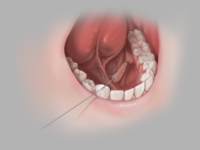
Gain and maintain access with a wire guide.
Sialendoscopy starts with accessing the salivary duct through the papilla. A flexible Cook soft-tipped wire guide can be maneuvered into the duct. This wire guide allows you to gain and maintain ductal access throughout the procedure, which reduces trauma and maximises efficiency.
 Learn More
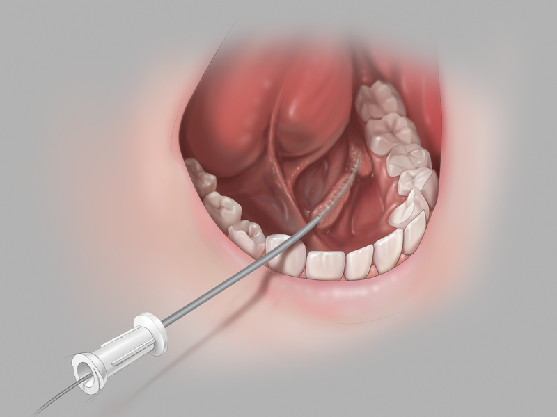
Minimise trauma with sequential dilation.
Once you establish access with the Cook soft-tipped wire guide, you can introduce a series of flexible dilators over the wire guide in order to expand the papilla and to prepare the salivary duct for the introduction of procedural instruments.
 Learn More
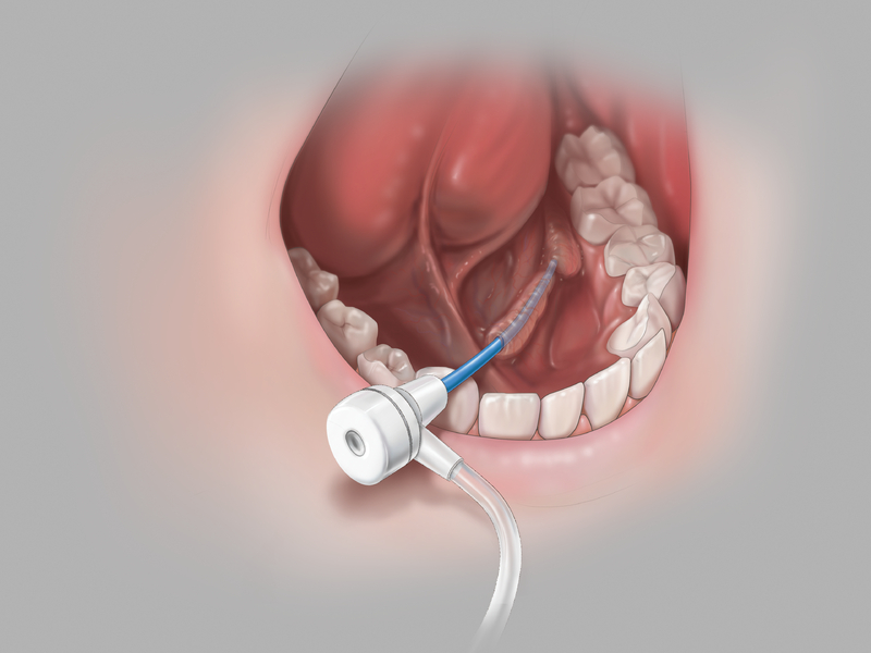
Create a true working channel.
After you expand the papilla with the dilator set, you can create an open working channel into the salivary duct by passing the sheath over the wire guide. The sheath also serves to protect the ductal wall as you insert and remove procedural instruments.
 Learn More
Manage challenging salivary stones with durable nitinol stone extractors.
Once you have gained access to the duct, you can insert the NCircle® or NGage® nitinol baskets into the duct to remove salivary stones and fragments. The NCircle® Tipless Salivary Stone Extractor has a tipless configuration that allows you to position the basket against the mucosal lining.
 Learn More
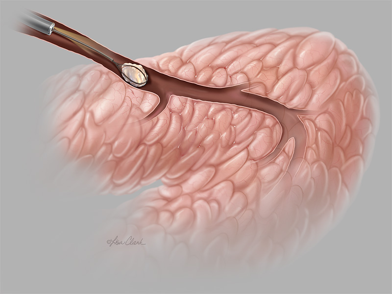
Manage challenging salivary stones with durable nitinol stone extractors.
Once you have gained access to the duct, you can insert the NCircle® or NGage® nitinol baskets into the duct to remove salivary stones and fragments. The NGage® Salivary Stone Extractor works as both a basket and grasper in one.
 Learn More
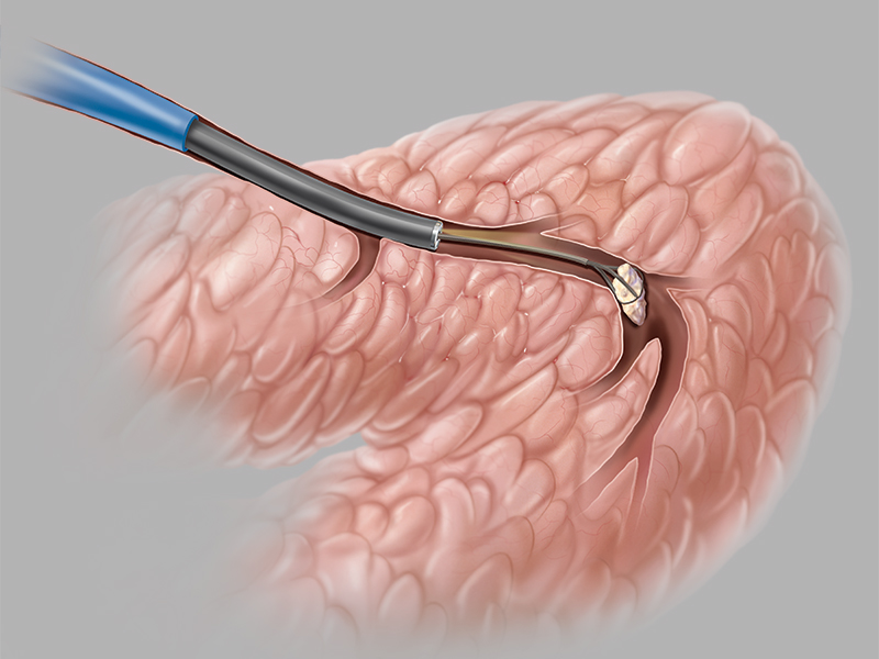
Expand your ability to treat obstructive salivary gland disorders.
This flexible, soft-tipped catheter is inserted over a wire guide or through the Kolenda Introducer Sheath to irrigate the submandibular or parotid salivary ducts or flush stone fragments.
 Learn More
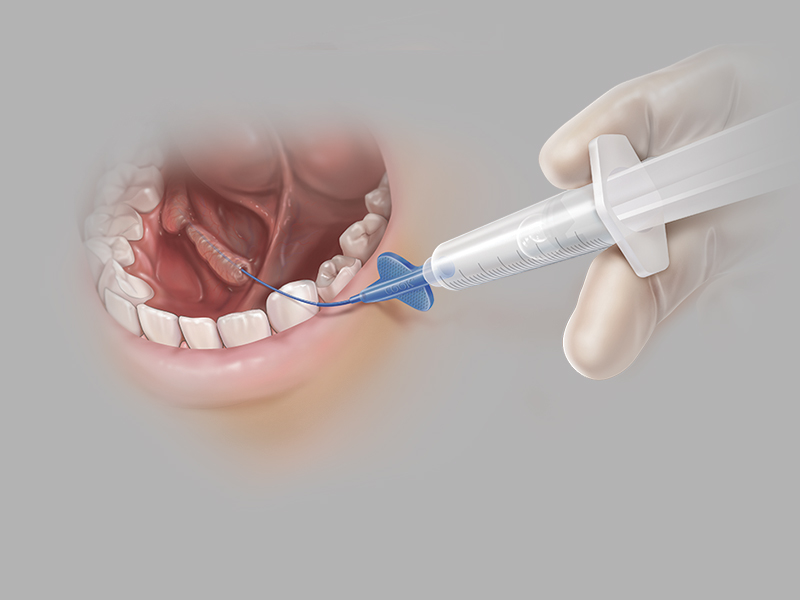
Accurately dilate salivary duct strictures.
Once you have created a working channel in the duct, you can insert the balloon catheter over a wire guide and use alongside a sialendoscope for direct visualisation to inflate the balloon at the stricture site.
 Learn More
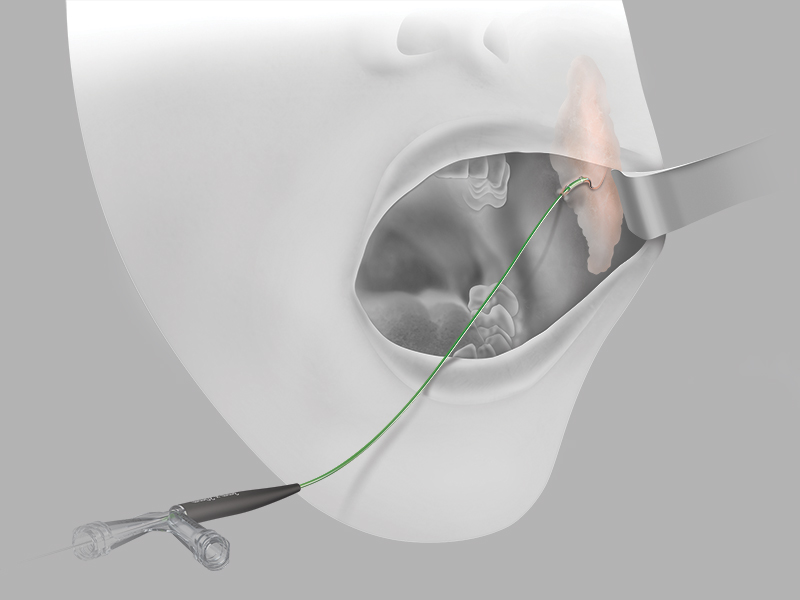
Advance® Salivary Duct Balloon Catheter Animation
Sialendoscopy Procedural Video
SialoCath™ Salivary Duct
Catheter Animation
Cook Medical is a proud supporter of several additional training opportunities. If you are interested in receiving information on future training opportunities, please visit our Vista website.
 View the Courses
 Contact Us
 For more information please contact us at CookOHNS@CookMedical.com with your name, location, and inquiry.
For more information about OHNS, visit cookmedical.eu.